
Before and After X-Rays — Disc Height Measurements
Disc height is one of the measurable changes we track during a course of DRX9000 care. The x-rays below are from four TriMed Spine & Joint patients, imaged before and after completing their DRX9000 protocol at our Boca Raton office.
Cervical spine
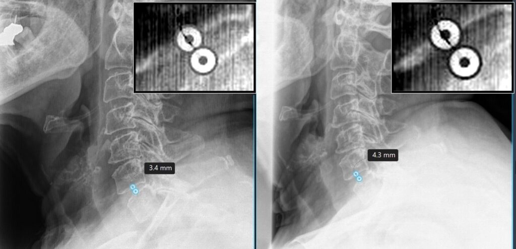
Case 1 — C6-C7: disc height changed from 3.4mm to 4.3mm (+0.9mm) over 7 months following the TriMed DRX9000 protocol.
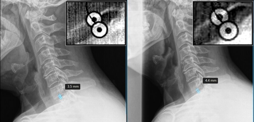
Case 2 — C7-T1: disc height changed from 3.5mm to 4.4mm (+0.9mm) over 3 months following the TriMed DRX9000 protocol.
Lumbar spine
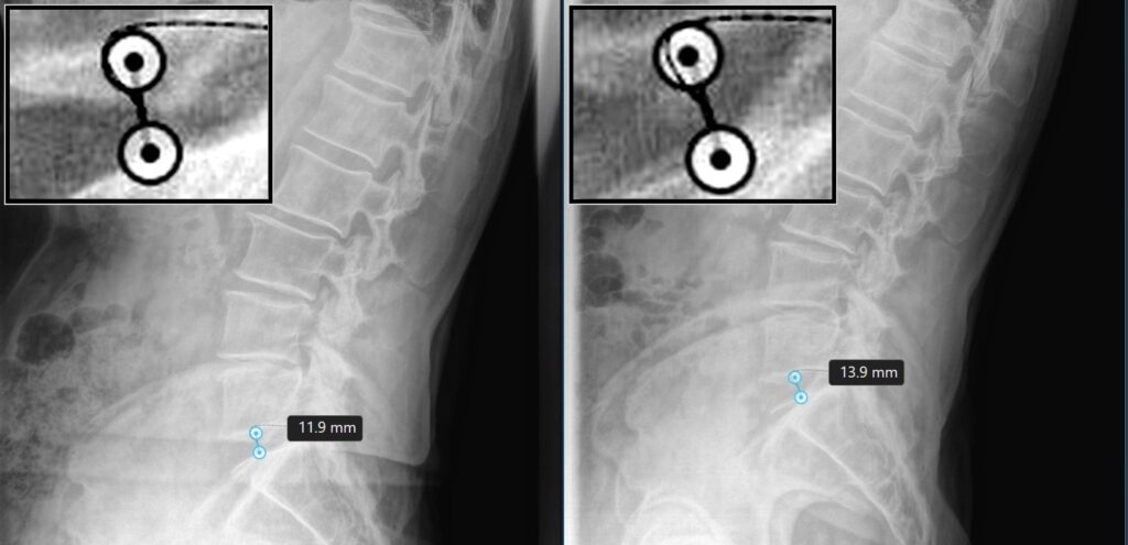
Case 3 — L5-S1: disc height changed from 11.9mm to 13.9mm (+2.0mm) over 2 months following the TriMed DRX9000 protocol.
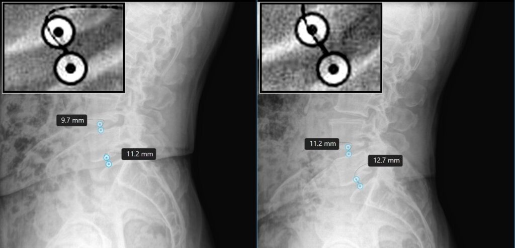
Case 4 — L4-L5 and L5-S1: L4-L5 disc height changed from 9.7mm to 11.2mm (+1.5mm). L5-S1 disc height changed from 11.2mm to 12.7mm (+1.5mm). Both changes occurred over 2.5 months following the TriMed DRX9000 protocol.
CASE EXAMPLES — INDIVIDUAL RESULTS VARY
These fully de-identified radiographs show individual patients before and after completion of a DRX9000® spinal decompression protocol. Intervertebral discs cannot be directly visualized on X-ray and are therefore better evaluated with MRI. Disc-height measurements shown are software-measured, specific to these images and individual cases, and should not be interpreted as expected results, since outcomes vary from case to case. Radiographic appearance and measurements may be affected by patient positioning, anatomy, condition severity, degenerative changes, treatment compliance, and other individual factors. No particular clinical or radiographic outcome is promised or guaranteed.
Read more about DRX9000 non-surgical spinal decompression in Boca Raton.
Pre- and Post-Treatment MRI Comparisons
EDUCATIONAL IMAGING — COURTESY EXCITE MEDICAL
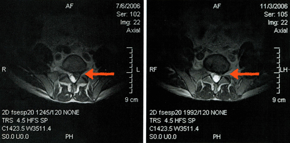
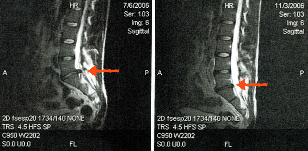
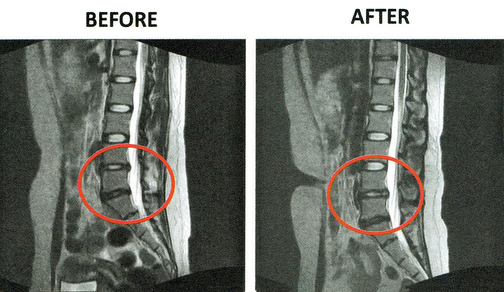
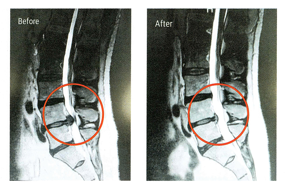
These MRI comparisons are educational images provided by Excite Medical, the manufacturer of the DRX9000®. They illustrate the type of disc changes documented in peer-reviewed research and do not depict TriMed Spine & Joint patients or images from the cited studies. Individual outcomes vary, and no particular clinical or imaging outcome is promised or guaranteed.

Why We Compare MRIs Before and After Treatment
An MRI is the clearest way to document what is happening to a disc. Comparing a scan taken before a course of DRX9000 spinal decompression therapy with one taken afterward lets a clinician show a patient exactly what changed rather than relying on how they say they feel. The MRI comparisons shown here are educational images courtesy of Excite Medical, illustrating the type of disc changes documented in peer-reviewed research.
What to Look For
When reviewing a pre- and post-treatment comparison, the findings that matter most are the position of the disc material relative to the spinal canal and nerve root, the height of the disc space between the two vertebral bodies, and the amount of space remaining around the nerve. Arrows on these images mark the level being compared.
We also track disc height on standing X-rays. You can see measured examples in our case write-ups, and read patient testimonials or our DRX9000 frequently asked questions.
What does an MRI show after spinal decompression?
A follow-up MRI or x-ray is read against the earlier study to see whether the disc material has changed position relative to the spinal canal and nerve root, whether the height of the disc space has changed, and how much room is left around the nerve. Those three findings are objective: they either changed or they did not, independent of how the patient reports feeling that week.
Can non-surgical spinal decompression increase disc height?
There is published evidence that it can. In a retrospective cohort of lumbar spine patients, non-surgical spinal decompression was associated with an increase in disc height on imaging (Apfel CC, Cakmakkaya OS, Martin W, et al. BMC Musculoskeletal Disorders. 2010;11:155). Measured before-and-after films from our own patients are published on our DRX9000 results page. Individual results vary and no outcome is guaranteed.
Does a change on my MRI mean my pain will improve?
Not necessarily. Imaging findings and symptoms do not always move together: some patients feel substantially better before their imaging changes, and some show a measurable change on film while still having symptoms that need further care. Imaging is one piece of evidence used alongside your examination and your own report of function.
Do I need an MRI before starting DRX9000 treatment?
Treatment decisions are based on a physical examination together with imaging of the affected level. If you already have a recent MRI, bring it or have the images sent to us before your visit. If you do not, Dr. Cantor will tell you what imaging is needed before decompression can start safely.
Are the images on this page from real patients?
The X-ray cases on this page are real TriMed Spine & Joint patients, imaged before and after completing their DRX9000® protocol at our Boca Raton office. The MRI comparisons are educational images provided by Excite Medical, the manufacturer of the DRX9000 — they illustrate the type of disc changes documented in peer-reviewed research and do not depict TriMed patients. Individual outcomes vary.
Individual Results May Vary
These images document individual cases and are not a prediction of what any other patient will experience. Whether spinal decompression is appropriate for you depends on your diagnosis, your imaging and your medical history. Call 1 (855) 487-4633 to arrange a review of your MRI with Dr. Cantor.